
Mole Mapping & Skin Cancer Screening
Early Detection • Digital Imaging • AI-Supported Dermatologic Follow-Up
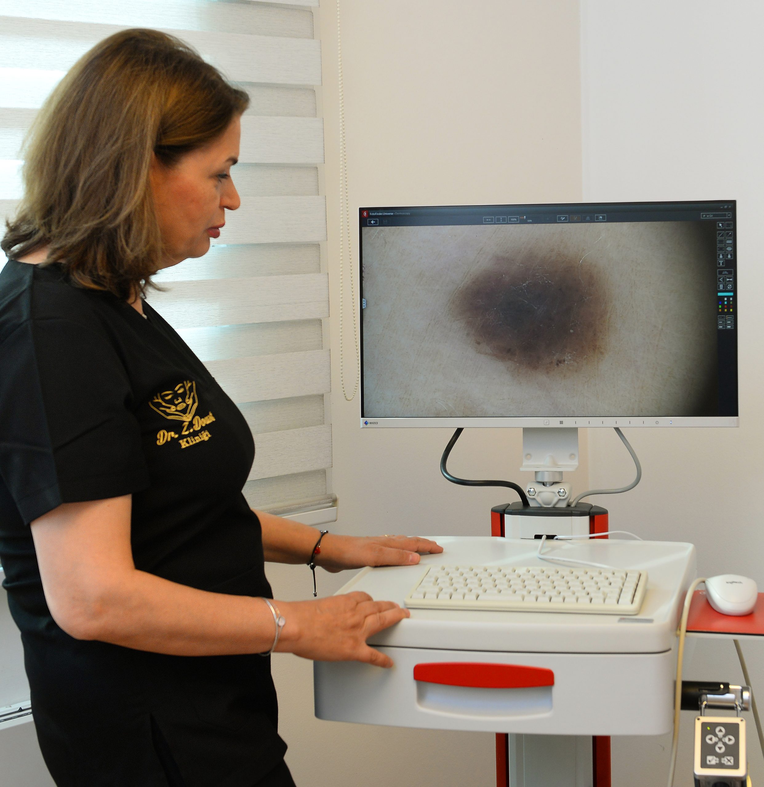
Mole mapping is an advanced dermatologic screening method used to monitor, document, and analyze moles with high-resolution cameras and computer-assisted systems. Through periodic follow-ups, even the smallest structural changes can be detected early, allowing timely intervention for suspicious lesions and reducing skin cancer risk.
What Is Mole Mapping?
Mole mapping involves photographing the entire body using specialized imaging systems. These images are analyzed with:
- High-resolution macro and dermoscopic cameras
- Computer-based comparison software
- Artificial intelligence risk evaluation tools
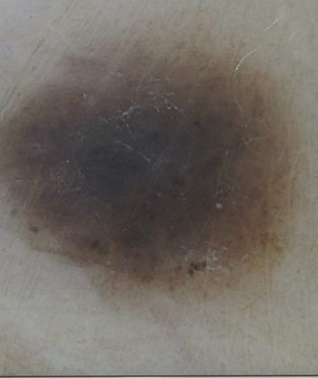
Each mole is examined for asymmetry, border irregularity, color changes, and size variations—criteria associated with early melanoma detection.
Who Should Have Mole Mapping?
- Individuals with many moles
- Those with atypical or dysplastic nevi
- Personal or family history of skin cancer
- Light skin, freckles, or sun-sensitive skin
- Anyone experiencing changes in existing moles
How Does the Process Work?
- Full-body imaging
- Digital dermoscopic examination
- AI-supported analysis & risk scoring
- Periodic comparisons every 6–12 months
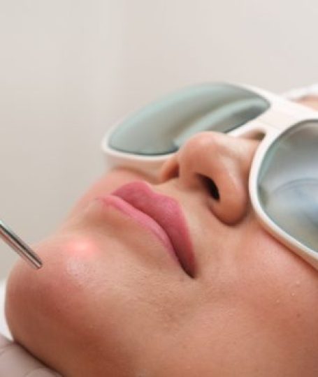
Treatment When Necessary
Suspicious lesions can be removed with minimal scarring using advanced surgical techniques.